4 Allergy and Immunology

4.1 Adverse Drug Reactions

4.1.1 Type A

  • Predictable, dose dependent (ex overdose, SEs, drug interactions. 85-95%

4.1.2 Type B

  • Unpredictable hypersensitivity reactions (intolerance, idiosyncracy, immunologic). 10-15%.

I

  • Immediate (mins-hrs)
  • IgE mediated
  • Anaphylaxis, angioedema, hives, hypotension, N/V/D

II

  • Delayed (variable)
  • Cytotoxic
  • Hemolysis, thrombocytopenia, neutropenia

III

  • Delayed (weeks)
  • Immune-complex
  • Serum sickness, arthus reaction, vasculitis

IV

  • Delayed (days to weeks)
  • Cell-mediated
  • Contact dermatitis, SJS/TEN, DRESS

4.1.3 Organ-specific ADRs

Type Manifestation Example drugs
Exanthems Diffuse fine macules/papules days after drug initiation Allopurinol, aminopenicillins, cephalosporins, AEDs, sulfonamides
Urticaria / Angioedema W/in minutes of drug initiation B-lactam antibiotics, ACEi
Fixed Eruption Hyperpigmented plaques that recur in same site Tetracyclines, NSAIDs, carbamazepine
Pustules Acneiform, Acute generalized eczematous pustulosis Steroids, sirolimus, antibiotics, CCBs
Bullous Tense or flaccid blisters Furosemide/vanco, capropril/penicillamine
SJS Fever, erosive stomatitis, ocular involvement, purpuric macules (face, trunk) w/ <10% epidermal detachment Sulfa antibiotics, AEDs, oxicam NSAIDs, and allopurinol
TEN Similar to SJS but w/ > 50% epidermal detachment Same as SJS, mortality as high as 50%
Lupus (skin) Erythematous / scaly plaques in photodistribution Hydrochlorothiazide, CCB, ACEis
Hematologic Hemolytic anemia, thrombocyto/granulocytopenia Penicillin, quinine, sulfonamides
Hepatic Hepatitis, cholestatic jaundice acetaminophen, sulfonamides
Pulmonary Pneumonitis, fibrosis Bleomycin, Nitrofurantoin, MTX
Renal Interstitial nephritis, MGN Penicillin, sulfonamides, allopurinol

4.1.4 Multiorgan ADRs

Type Manifestation Example drugs
Anaphylaxis Urticaria/angioedema, bronchospasm, GI sx, hypoTN B-lactam antibiotics, monoclonal Abs
DRESS Cutan. eruption, fever, eosinophilia, hep. dysfunction, LAD AEDs, sulfonamides, minocycline, allopurinol
Serum Sickness Urticaria, morbiliform rash, arthralgias, fever Heterologous abs, infliximab, bactrim, PCN
SLE Arthralgias, myalgias, fever, malaise Hydralazine, Procainamide, Isoniazid
Vasculitis Cutaneous or visceral vasculitis Hydralazine, penicillamine, propylthiouracil

Desensitization Definition: give increasing doses over hours

  • Mast cells and basophils unreactive to Ag activation (only for Type I HSRs)

Result: Temporary tolerance

  • patient can receive the drug at usual intervals. When drug is stopped, desensitization ends (d-wk)

4.2 Anaphylaxis

Definition Acute, life threatening systemic HSR (min-hrs) w/ >= 1/3 of the following criteria:

Hives plus another system: acute onset illness (mins-hrs) involving skin/mucosa, or both, and >= 1 of the following: respiratory compromise, reduced BP or symptoms of end-organ dysfunction

Two systems involved: >= 2 of the following must occur rapidly after exposure to a likely allergen (mins-hrs): skin-mucosal involvement, respiratory compromise, reduced BP or associated symptoms of end-organ dysfunction, persistent GI symptoms

Hypotension: reduced BP after exposure to known allergen (mins-hrs)

4.2.1 Allergens

  • Meds (B-lactams, ASA/NSAIDs), food, insects, cold/heat, exercise, latex

4.2.2 Clinical

  • Skin involvement in 90%, respiratory in 70%, CV (hypotension) in 45%, GI in 45%
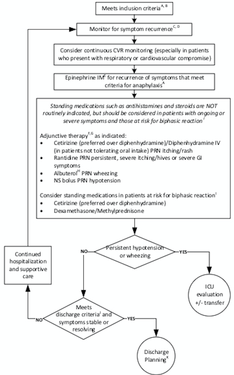
  • Monitor for biphasic reaction (4-25% occurrence)
  • sx recur w/in 10h (but up to 72h)

4.2.3 Severe Reaction

  • Hypotension w/ wide PP, AMS/confusion, syncope, cyanosis, dyspnea, hypoxia

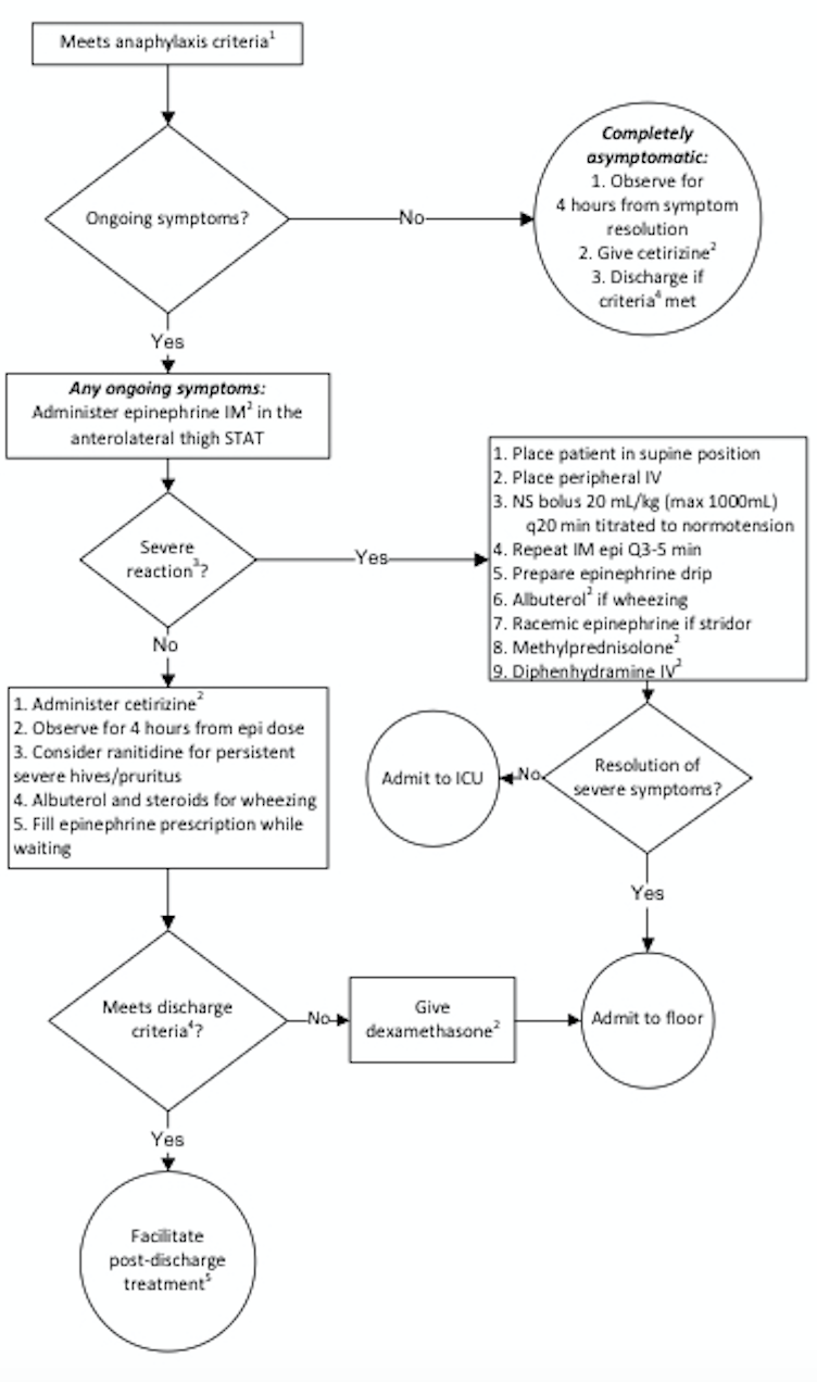
4.2.4 Management Emergent

Inpatient

4.2.5 Med Dosing

  • Epinephrine IM (1 mg/mL)
  • 0.01 mg/kg (<10 kg), 0.15 mg/kg (10-25kg), 0.3 (>25 kg)
  • Cetirizine - 2.5 mg (6mo-2 yrs), 5 mg (2-5 yrs), 10 mg (≥6 yrs)
  • Diphenhydramine - 1 mg/kg IV/PO (max 50 mg)
  • Dexamethasone 0.6 mg/kg (max 16 mg) OR methylprednisolone 1 mg/kg (max 60)
  • Ranitidine - 2 mg/kg PO (max 150 mg) OR 1 mg/kg IV (max 60 mg)

4.2.6 ED Discharge Criteria

No hypotension, resolved wheezing, ≤ 2 doses of Epi

4.2.7 Post-discharge Treatment

3 days of Cetirizine daily, consider ranitidine, f/u with PCP/Allergy

4.3 Primary Immunodeficiencies

4.3.1 Pathophysiology

  • Genetic defects in the adaptive (B- or T-cell) or innate (phagocytes, complement) immune systems lead to recurrent infections
  • Over 200 distinct disorders: B cell defects (65%), combined B and T cell deficiencies (15%), phagocytic disorders (10%), T cell deficiencies (5%), and complement deficiencies/others (5%)

4.3.2 Epidemiology

The overall incidence is 1:10000, and overall prevalence is 1:2000.

Clinical Can be nonspecific and broad

Constitutional: Poor growth, failure to thrive

GI: chronic diarrhea.

Derm: Atopic and non-atopic dermatitis, severe diaper rash, neonatal rash, anhydrosis, as well as delayed separation of the umbilical cord (LAD)

Immuno: Recurrent infections, autoimmunity Family history of consanguinity or family history of immunodeficiency or unexplained childhood deaths puts a child at higher risk of having or developing a primary immunodeficiency

4.3.3 Physical Exam

Vital signs: Growth parameters

General exam: Note dysmorphisms, including teeth and hair (abnormal in NEMO). Look for infectious sources (sinusitis, otitis, pneumonia, thrush, diaper rash)

HEENT exam: Note tonsils (absent in XLA) and examine for thrush and other signs of infection such as sinusitis or recurrent otitis media

CV exam: Note any cardiac anomalies including heart sounds, pulses, perfusion, and overall volume status as cardiac anomalies can be a part of certain syndromes associated w/ immunodeficiency syndromes (e.g.: DiGeorge Syndrome)

Respiratory: Note symmetry of lung exam, quality of air entry, and lung sounds as pulmonary anomalies may be a manifestation of immunodeficiency syndromes

GI: A thorough GI exam including abdominal exam for elements like hepatosplenomegaly and rectal exam for possible anal atresia is important

GU: Primary immunodeficiencies can also lead to GU anomalies; assess for absence/presence of appropriate male/female organs in the correct number

Derm exam: Skin exam for eczema/dermatitis (i.e. WAS, SCID, hyper IgE syndrome) as well as erythroderma (Omenn Syndrome). Note telangiectasia (AT), warts, granulomas, poor wound healing or ulcers

Neuro: A thorough neuro exam may also hint at the etiology of an immunodeficiency (ataxia-telangiectasia), an infection such as meningitis, or may help elucidate an alternate cause of symptoms

4.3.4 Diagnosis

Initial labs: CBC w/ differential (note especially lymphopenia), chem7, albumin, urinalysis, ESR, CRP, quantitative immunoglobulins (IgG, IgA, IgM, IgE), specific vaccine antibody studies (tetanus, HiB, pneumococcal).

Follow-up labs: HIV testing. B- and T-cell subset, complement screening (C3, C4, AH50, CH50), vaccine challenge (administer pneumococcal or other vaccine and measure titers 4-6 weeks later), Dihydrorhodamine (DHR) assay (CGD). Leukocyte adhesion defect testing (LAD).

Advanced lab analysis: T cell proliferation studies (mitogen, antigen), T and B cell memory panels, NK cell function assays, Toll-like receptor assays. Immunodeficiency genetic panel. Whole exome or whole genome sequencing.

4.3.5 Treatment

Varies widely based upon the deficiency. Common therapies include prophylactic antibiotics, IVIG, bone marrow transplant.
4.4 Indications for a Primary Immunodeficiency Evaluation

  • ≥8 ear infections w/i one year
  • >2 serious sinus infections w/i one year
  • >2 pneumonias w/i one year - FTT, poor weight gain, or abnormal growth parameters
  • Abnormal physical exam suggestive of syndrome
  • Recurrent skin or organ abscesses
  • Persistent thrush (mouth/skin), at >12 months of age
  • Severe or overwhelming infection
  • Infection w/ unusual organisms
  • Need for intravenous antibiotics to clear infections
  • Infections w/ opportunistic organisms (Aspergillus, Pneumocystis)
  • Severe forms of viral infections (HSV, VZV, EBV)
  • Complications from a live vaccine
  • A family history of primary immunodeficiency
  • Abn. TRECs on newborn screen x2
  • Abn. screening CBC (profound leukopenia, lymphopenia, eosinophilia)

4.5 Diagnostic Approach to Primary Immunodeficiencies

4.5.1 Initial Labs (most cases)

  • CBC w/ differential - Quantitative immunolgobulins (IgG, IgA, IgM, IgE)

  • Specific antibody studies (tetanus, HiB (PRP), pneumococcal)

4.5.2 Next Step

(include w/ initial labs if suspicious of specific disorder)

  • B- and T-cell subsets

  • T cell proliferation studies (mitogen, antigen)

  • Complement screening (CH50, AH50, C3, C4)

  • DHR (dihydrorhodamine assay for CGD)

4.5.3 Advanced (Depending on specific history)

  • T and B cell memory panels
  • NK cell function assay
  • Toll-like receptor studies
  • Specific genetic testing

4.6 Classification of Primary Immunodeficiencies

4.6.1 B-cell (Humoral): decreased B-cell numbers and/or impaired antibody production

Diseases

  • X-linked agammaglobulinemia
  • Transient hypogammaglobulinemia of infancy
  • IgA or IgG selective Ig deficiency

Clinical Manifestations

  • Presents <12 mo old (3-6 mo, due to loss of maternal antibody)
  • Bacterial infxn (sinusitis, otitis, pneumonia)
  • Chronic diarrhea, FTT - Bronchiectasis
  • Enteroviral meningoencephalitis (chronic)

Organisms

  • Encapsulated: S. pneumo, HiB, N. meningitidis, S. typhi
  • GI: Giardia, Campylobacter
  • Also: S. Aureus, Pseudomonas, Enteroviral meningoencephalitis

Vaccine Issues

Do not give live vaccines for severe defects. Vaccination is not necessary if on IgG replacement. Effectiveness of other vaccines is uncertain

4.6.2 T-cell Defects

(Cellular): lack of or decreased number of T-cells

Diseases

DiGeorge Syndrome, SCID (T-/B+)

Clinical Manifestations

  • Presents at birth/early infancy
  • Mucocutaneous candidiasis
  • Severe viral infections
  • Bacterial, fungal, opportunistic infections
  • Warts or severe eczema
  • Chronic diarrhea, FTT

Organisms

  • Candida, PJP, Mycobacterium, S. typhi
  • VZV, HSV, CMV

Vaccine Issues

Do not give live virus vaccines if substantial T cell defect

4.6.3 Combined B/T Cell Defect

4.6.3.1 Diseases

  • SCID (T-/B-)
  • CVID
  • Wiskott-Aldrich syndrome
  • Ataxia-telangiectasia
  • X-linked lymphoproliferative disease (XLP)
  • Hyper IgE syndrome
  • DOCK8 deficiency
  • ZAP70 deficiency

4.6.3.2 Clinical Manifestations

  • Presents during 1st year of life. XLP/CVID can present as teens/adults
  • Infections (sinusitis, otitis, pneumonia)
  • Abscesses (recurrent)
  • Chronic diarrhea, gastroenteritis, FTT
  • Mucocutaneous candidiasis
  • Viral/opportunistic/fungal infections
  • Increased cancer risk

Organisms

  • Candida, PJP, Mycobacterium, encapsulated bacteria

  • VZV, HSV, CMV infections

Vaccine Issues

Do not give live vaccines (OPV, BCG, smallpox, YF, live influenza, MMR, MMRV, rotavirus). Effectiveness of other vaccines is uncertain.

4.6.4 Phagocytic Defects

4.6.4.1 Diseases

  • Chronic granulomatous disease (CGD)
  • Leukocyte adhesion deficiency (LAD)
  • Chediak-Higashi syndrome (CHS)

4.6.4.2 Clinical Manifestations

  • Typically presents in infancy
  • Poor wound healing
  • Delayed separation of the umbilical cord (LAD)
  • Lymphadenitis/abscesses
  • Catalase (+) bacterial infections (CGD)
  • Candidiasis
  • Chronic gingivitis, oral disease
  • Hepatosplenomegaly

4.6.4.3 Organisms

  • Catalase (+) bacteria:
    • S aureus
    • Pseudomonas
    • Burkholderia cepacia
    • Nocardia
    • Enterobacteria erratia and Klebsiella
  • Fungal infections:
    • Aspergillus
    • Candida albicans

4.6.4.4 Vaccine Issues

  • Live viral vaccines contraindicated in CH & LAD, but OK in CGD
  • Live bacterial vaccines are contraindicated. Other vaccines are safe/ effective

4.6.5 Complement Defects

4.6.5.1 Diseases Classical pathway:

  • C1q, Cqr, C1s, C2, C4
  • Hereditary angioedema (C1-est)
  • C2: most common in Causasians

Lectin pathway: - MBL, M-/L-/H-ficolin, CL-11, MASPs

Alternative pathway: - Factors D, B, and properdin

4.6.5.2 Clinical Manifestations

  • Can present at any age
  • Angioedema of the face, lips, hands, feet, GI tract, throat (C1-inh)
  • Recurrent sinopulmonary infections
  • Bacteremia/pyogenic bacterial infections
  • Meningitis - Autoimmune disease (lupus-like)
  • Often autosomal dominant inheritance
  • Associated w/ atypical HUS

4.6.5.3 Organisms Encapsulated bacteria, Neisseria

4.6.5.4 Vaccine Issues

  • No vaccine contraindications
  • Refer to CDC guidelines re: additional vaccinations for protection against encapsulated bacteria

4.7 Selected Primary Immunodeficiencies

Disorder Cell Gene Age Presentation Labs
Chediak-Higashi Syndrome (CHS) Phagocyte CHS1, AR Infancy -Oculocutaneous albinism, recurrent pyogenic infections (S. aureus) -May present with accelerated phase or HLH: fever, jaundice, hepatosplenomeglay, lymphadenopathy, bleeding, neurologic changes -Lysosomes unable to fuse with phagosomes to lyse bacteria -Neutropenia -Giant lysosomal granules in neutrophils -Impaired T/NK cell function
Chronic Granulomatous Disease (CGD) Phagocyte Multiple phagocyte oxidases (gp91phox), X-linked/AR Infancy-adult, most <5yo -Recurrent bacterial/fungal infections, often w/ encapsulated and catalase-positive organisms -Granulomas and cold abscesses, both superficial and deep -Inability of neutrophils to generate oxidative burst, but chemotaxis and phagocytic function intact -Normal neutrophil count -Reduced superoxide production when stimulated in vitro -DHR assay
Selective IgA Deficiency B-cell - >4yo -Most patients (85-90%) are asymptomatic -Recurrent sinopulmonary infection (H. influenzae, S. pneumo), Giardia lamblia infections, autoimmune disease -Increased risk of anaphylaxis to blood products -Low IgA, normal IgG/M
X-linked Agammaglobulinemia (XLA) Complete absence of mature B-cells BTK, X-linked recessive 3-18mo -Recurrent bacterial infections: sinuses, ear, lung (S. pneumo, HIB, S. pyogenes, Pseudomonas) -Exam: absent tonsils and adenoids -Low levels of IgG, IgM, IgA -Reduced/absent CD19/20 B-cells
DiGeorge Syndrome Normal to severe T-cell immunodef Del. 22q11.2 & 10p13-14 Infancy -Triad: hypoplastic thymus, conotruncal cardiac/aortic arch defects, hypoparathyroidism -Characteristic faces: low set ears, ocular hypertelorism, bulbous nasal tip -Hypocalcemia -Reduced CD3+ T cells -Abnormal cardiac echo
Ataxia Telangiectasia (AT) B- and T-cell ATM, AR >1yo -Progressive cerebellar ataxia, oculocutaneous telangiectasia, diminished/absent deep tendon reflexes -Recurrent sinopulmonary infections -Increased risk of malignancy -Selective IgA deficiency -Low T-cell numbers -Elevated serum AFP
Common Variable Immunodeficiency (CVID) Impaired T-cell function, B-cell maturation - Childhood-Adolescence -Recurrent sinopulmonary infections, autoimmunity, chronic lung disease -Poor response to protein, polysaccharide vaccines (tetanus, PCV) -Significantly reduced IgG -Reduced IgA and/or IgM
Hyper IgE Syndrome B- and T-cell STAT3, AD First wks of life -Papulopustular rash, skin abscesses (S. aureus), eczema, retained primary teeth -Coarse/thickened facial features, frontal bossing, wide alar base of nose -Eosinophilia -Elevated IgE
Severe Combined Immunodeficiency (SCID) B- and T-cell, depending on the type Multiple (RAG1, RAG2, ADA, Artemis, IL2RG) Part of newborn screen -Persistent mucocutaneous candidiasis, FTT, recurrent fevers, chronic diarrhea -Infections with adenovirus/CMV/EBV/RSV can be fatal -Live-attenuated vaccines can be fatal -Immunologic emergency: positive pressure room, urgent work-up and evaluation for bone marrow transplant -NBS: low TRECs -CXR: absence of thymic shadow -Absolute T-cell count <300, abnormal T-cell proliferation studies, presence of maternal T-cells in circulation

4.8 Selected Immunodeficiencies

Disorder Cell Gene Age Presentation Labs
Wiskott-Aldrich Syndrome B- and T-cell WAS X-linked Infancy -Triad: thrombocytopenia (small platelets), eczema, immunoglobulin abnormalities -Chronic otitis media/sinusitis, infection with encapsulated organisms -IgG nml, IgM low, IgA/E elevated -Decreased number of T cells -Thrombocytopenia
X-linked Lymphoproliferative Disease (XLP) T- and B-cell XLP/SH2D 1A, X-linked R Childhood -Fulminant EBV infection (often w/ hepatitis, hepatosplenomegaly, liver failure), often causing secondary hemophagocytic lymphohistiocytosis or aplastic anemia -Inc risk of malignancy, especially lymphoma -B/T cell numbers normal; function is abnormal -IgG is low, IgM is increased -Anemia, thrombocytopenia

4.9 Specific Antibody Deficiencies

Presentation IgG IgA IgM IgG subclass Vaccine response B cells
Subclass deficiency Recurrent severe infections (controversial) NL NL NL At least 1 is low LOW NL
Selective IgA Deficiency Asymptomatic or associated w/ autoimmune, GI, atopic disorders NL LOW NL NL NL OR LOW NL
Hyper IgM Syndrome Severe infections, including PJP LOW LOW NL OR HIGH LOW LOW NL
Specific Antibody Deficiency Often asymptomatic, inadequate antibody response to polysaccharide antigen NL NL NL NL LOW NL
CVID Recurrent infections LOW NL OR LOW NL OR LOW LOW LOW NL

4.10 Characteristics of Selected SCID disorders

4.10.1 T-, B+, SCID

4.10.1.1 Gene defects

  • IL2RG (most common form, X-linked)
  • JAK3
  • IL7RA
  • IL2RA
  • CD3D/E/Z
  • PTPRC
  • CORO1A
  • ZAP70

4.10.1.2 Treatment Bone marrow transplant or gene therapy (IL2RG)

4.10.2 T-, B-, SCID

4.10.2.1 Gene defects

  • RAG1/RAG2 (common)
  • Artemis (common)
  • Adenosine deaminase (ADA, common)
  • PRKDC
  • AK2
  • LIG4
  • Cernunnos (NHEJ1)

4.10.2.2 Treatment

  • Bone marrow transplant or gene therapy (ADA)

  • ADA can be treated w/ gene therapy or enzyme replacement

4.11 General Approach to the Immunodeficient Patient

Approaching the immunodeficient patient in clinic:

  • See indications for testing/tests above.
  • Is antibiotic prophylaxis indicated?
  • Can they receive immunizations? If they can, have they mounted a sufficient immune response to vaccines (ie. do they need vaccine titers)?
  • Low threshold for antibiotic use in the event that infection is suspected.

Managing a sick immunodeficient patient:

  • Obtain blood culture and labs.
  • Give antibiotics/antivirals promptly.
  • Determine whether imaging/surgery is indicated (ie. drain an abscess).
  • Be aware of blood products given. Blood may need to be from CMV negative donors, filtered to remove WBCs, and irradiated.
  • Replace missing immune components (ie. IVIG)