8 Dermatology

8.1 Key Questions for Taking a Dermatologic HIstory

  • When did it start? (ask for timepoints, i.e., “before Christmas?”)
  • Does it itch, burn, or hurt?
  • When was the first episode?
  • Where on the body did it start? Was it present at birth?
  • How has it spread (pattern of spread)?
  • How have individual lesions changed? (Size, shape, color, itch, bleeding?)
  • Is there a family history? (eczema, acne, psoriasis, autoimmune disease)
  • What has made it worse or triggered it?
  • What have you tried for it? Did it help?
  • Hx of atopic triad (asthma, allergies, atopic dermatitis)?
  • New exposures: Medications? (Look at external med rec) Travel? Environmental?
  • Ask if they brought the medications they are using

8.2 Describing Dermatologic Lesions

8.2.1 Primary Lesion

Description of the “family” of a lesion Appearance
Macule Flat, not palpable; color change; <1cm (e.g., freckle, labial macule)
Patch Flat, not palpable; color change; >1cm (e.g., congenital nevus or large birthmark)
Papule Raised; <1 cm (e.g. mole, acne)
Plaque Raised; >1 cm usual flat topped (e.g. psoriasis)
Nodule Raised; round-topped lesion w/ depth; >0.5cm up to 1 cm (e.g. acne)
Tumor Very large, round-topped lesion w/ depth, exophytic, endophytic or level w/ skin surface ; >1cm (e.g. strawberry hemangioma of infancy)
Wheal Edematous, raised, hive-like
Vesicle Small fluid-containing blister <1cm (e.g. chickenpox, shingles zoster)
Bulla Large fluid-containing blister > 1cm (e.g. bullous pemphigoid)
Pustule Exudate filled; <1cm; can develop into furuncle, then abscess (e.g. pustular psoriasis)
Telangiectasia Dilated superficial capillaries
Verrucae Soft, tan-colored, cauliflower-like papules (e.g. warts caused by HPV)

8.2.2 Secondary Changes

Describe the changes that occur superimposed upon the primary lesion

  • Scale: Flakes of “dead skin”; thickening of outermost layer (stratum corneum)
  • Crust: Adherent, dried serum, exudate or blood on the skin
  • Desquamation: Thicker scale that is shedding off
  • Erosion: Loss of superficial layers of skin (epidermis only involved, does not scar); erosions lead to ulcerations
  • Ulceration: Loss of deeper layers of skin (extends to dermis, scars)
  • Fissure: Deep linear cracks in skin
  • Atrophy: Thinning of skin; can be in the epidermis, dermis, or subcutaneous fat
  • Excoriation: Erosions due to scratching
  • Lichenification: Thickened, leather-like skin, normal skin lines accentuated
  • Scar/keloid: Permanent fibrotic changes that result from damage extending into the dermis, keloids extend beyond the borders of the original defect
  • Umbilicated: Centrally indented

8.2.3 Color Descriptor

  • Erythematous: Blanchable red or pink hue in the skin, indicates vascular dilation; erythema is not a color per se
  • Purpuric: Violaceous color due to blood pigment
  • Petechial: Pinpoint, non-blanching bleeding into the skin from capillaries
    • Purpura = 1cm, ecchymoses > 1cm
  • Hyperpigmentation: Darker than normal skin color
  • Hypopigmentation: Lighter than normal skin color

8.2.4 Lesion Shapes

  • Annular: Ring-shaped lesion
  • Nummular/discoid/round: Coin-shaped lesion
  • Arcuate: Arc-shaped lesion
  • Linear Forming a line
  • Serpiginous: Wavy like a snake
  • Figurate/polycyclic: Lots of different ring shapes, rings within rings
  • Retiform/reticulate: Web or net-like, following vasculature
  • Targetoid: Lesions with a bull’s eye, or concentric rings or varying colors

8.2.5 Distribution

  • Clustered (agminated): Grouped together in a bunch but not running together
  • Discrete/scattered: Lesions are separate from one another
  • Acral: Over distal portions of limbs: finger tips, knuckles, elbows, knees, buttocks, toes, heels
  • Generalized: Throughout body
  • Photodistributed: Sun-exposed areas
  • Coalescing:

8.3 Newborn Derm

8.3.1 Neonatal Rashes

Condition Description Appearance
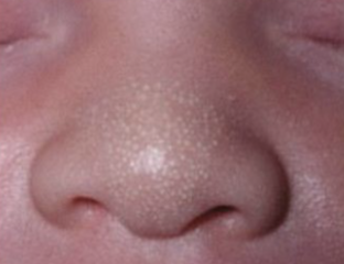
Sebaceous hyperplasia Minute, profuse yellow-white papules frequently on forehead, nose, lip, and cheeks
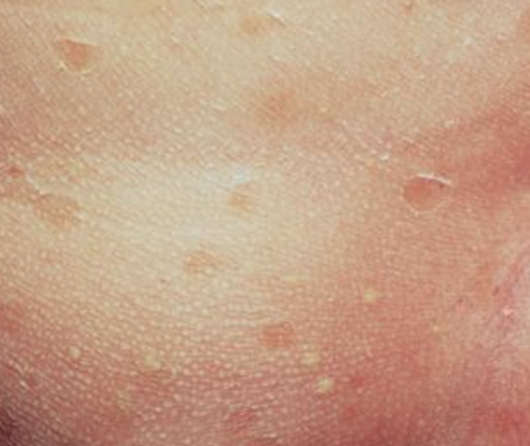
Milia/miliaria 1-2 mm pearly, opalescent cysts
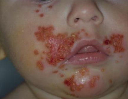
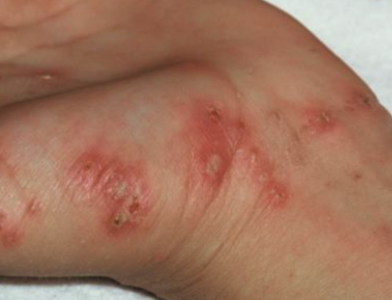
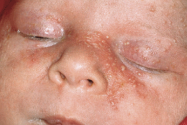
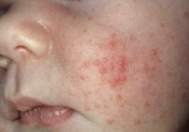
Neonatal acne (neonatal cephalic pustulosis) Inflammatory papules and pustules usually w/o comedonal lesions
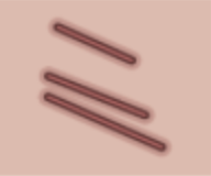
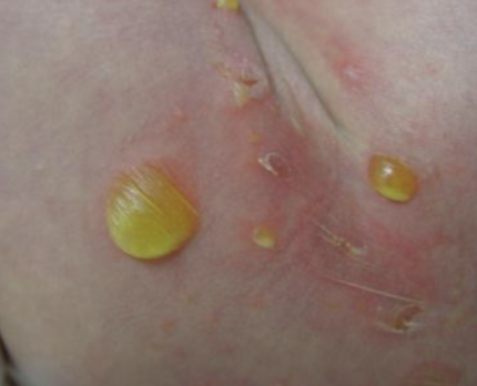
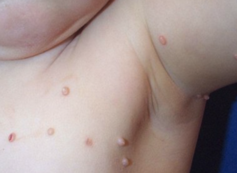
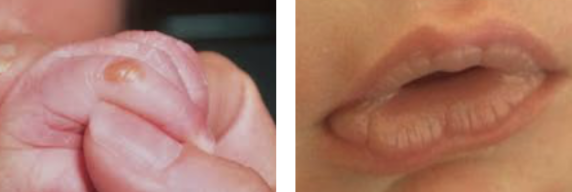
Sucking blisters Solitary or scattered superficial bullae on upper limbs of infants at birth (presumed in utero sucking)
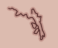
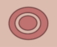
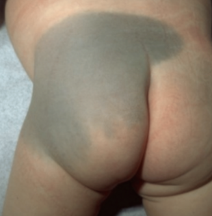
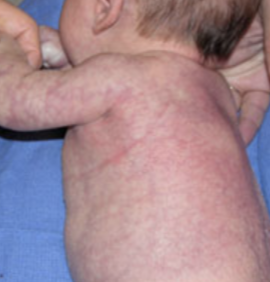
Cutis marmorata Evanescent, lacy, reticulated red and/or blue cutaneous pattern when exposed to low environmental temperatures
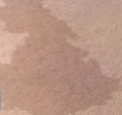
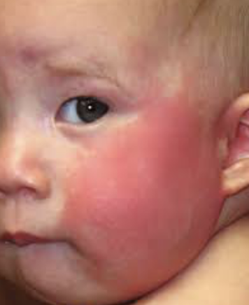
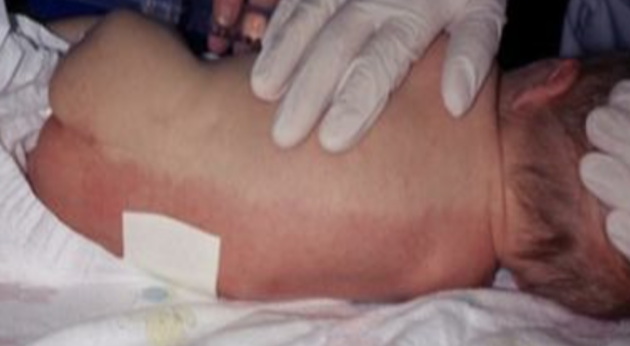
Harlequin color change When infant (usually immediate newborn period and in low birth weight infants) is laying on side, dependent area is deep red and upper half (longitudinally) is pale
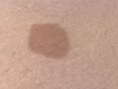
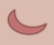
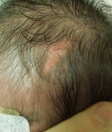
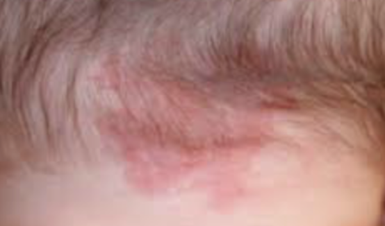
Nevus simplex (“salmon patch,” “stork bite,” “angel’s kiss”) Small, pink, ill-defined vascular macule usually on glabella, eyelids, upper lip and nuchal area
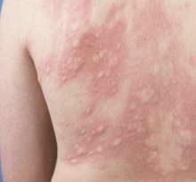
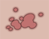
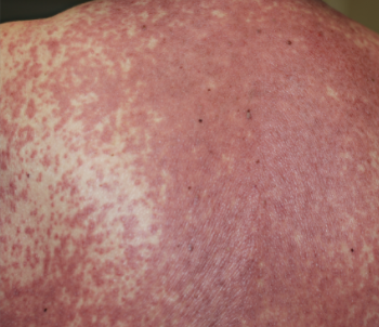
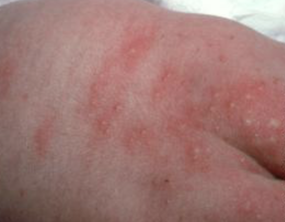
Erythema toxicum neonatorum (e tox) Benign, self-limited evanescent eruption usually in term infants presenting w/ firm, yellow-white papules and pustules w/ a surrounding erythematous flare; palms and soles are almost never affected
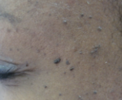
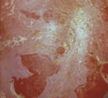
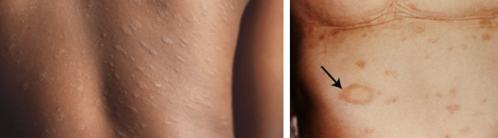
Transient neonatal pustular melanosis (TNPM) Superficial pustules, ruptured pustules w/ a fine scale, and hyperpigmented macules
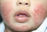
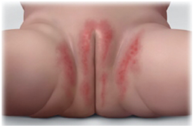
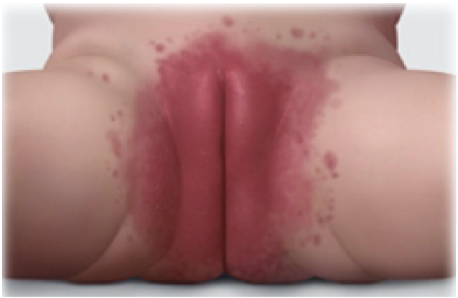
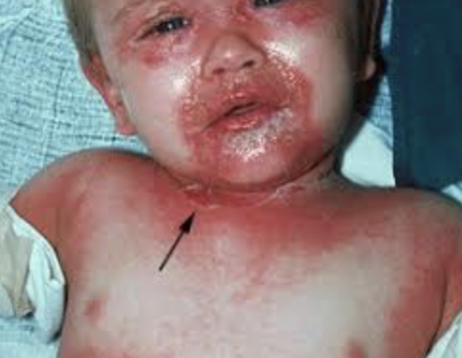
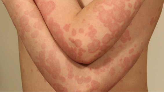
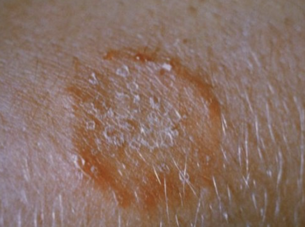
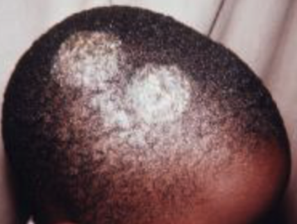
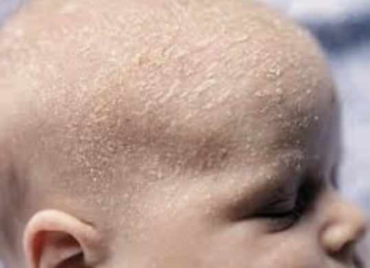
Seborrheic dermatitis Erythema and greasy scales usually on the scalp (“cradle cap”), face, forehead, trunk, intertriginous and flexural areas including diaper

8.3.2 Neonatal Birthmarks

Condition Description Appearance
Congenital melanocytic nevus (CNM, moles) - Often benign neoplasms composed of melanocytes
- Small and medium sized CMN have less than 1% risk of malignant transformation; large and giant lesions the risk is higher, ranging from 0-7.6%
Nevus sebaceous (organoid hamartoma) - Overgrown epidermis, sebaceous glands, hair follicles, apocrine glands and connective tissue that occurs primarily on scalp or face
- Presents as solitary, smooth, yellow-orange hairless patch, often oval or linear. Often becomes more pronounced in adolescence, appearing bumpy, warty, scaly.
Aplastic cutis congenita - Absence of skin present at birth that can be localized or widespread
- Can be an isolated finding or associated with other developmental anomalies
- Large scalp defects should be imaged to r/o underlying bone, vascular, or soft tissue defects
Congenital dermal melanocytosis (CDM; slate gray patch, Mongolian spot) Blue or slate-gray macular lesions
- Important to always point out to parents and to counsel them to point out to caretakers / daycare, as they can be mistaken for bruises
Vascular tumors - Infantile hemangioma, congenital hemangioma, pyogenic granuloma
- Hemangioma red flags: Beard distribution (evaluate airway), periocular (ophtho), paraspinal midline, hemangiomatosis (multiple small hemangiomas → evaluate for parenchymal hemangiomas, especially hepatic and CNS), very large hemangioma, associated thrill or bruit, head tilting
Vascular malformations Capillary malformation (nevus flammeus/Port wine stain), lymphatic malformations, cutis marmorata telangiectatica congenita (CMTC)

8.3.3 Diaper Dermatitis

Contact/Irritant Dermatitis Candida Dermatitis
Epidemiology Most common cause 2nd most common cause
Physical Exam Spares creases/skin folds “Beefy” red rash involving skin folds w/ satellite lesions
Management Topical barrier ointment/paste (petrolatum, zinc oxide) Topical antifungal (nystatin)

8.4 Dermatologic Emergencies

8.4.1 Stevens Johnson Syndrome (SJS)

8.4.1.1 Definition

Skin + 2 or more mucosa. 10-30% BSA.

8.4.1.2 Etiology

Infection & meds (sulfonamides anticonvulsants, NSAIDs, allopurinol, dapsone)

8.4.1.3 Presentation

Mucosal involvement, prodromal fever, sore throat, HA, malaise, erythematous target like lesions forming blisters that rupture

8.4.1.4 Management

  • Treat/discontinue underlying cause
  • Magic mouthwash for stomatitis, artificial tears for ocular involvement
  • Care to avoid scarring and adhesions
  • Hospitalize, treat like a burn patient (fluids, electrolytes, pain, prevent infection)

8.4.2 Toxic Epidermal Necrolysis (TEN)

8.4.2.1 Definition

Skin + 2 or more mucosa. >30% BSA.

8.4.2.2 Etiology

As above in SJS

8.4.2.3 Presentation

Extensive skin and mucosal involvement (conjunctival, oral, genital, pulmonary), large bullae that rupture and leave large erosions (Nikosky +)

8.4.2.4 Management

  • See SJS management above
  • Consider IVIG

8.4.3 Drug Reaction with Eosinophilis and Systemic Symptoms (DRESS)

8.4.3.1 Definition

Potentially life-threatening adverse drug-induced reaction characterized by skin rash, hypereosinophilia, liver involvement, fever, and lymphadenopathy

8.4.3.2 Etiology

Meds (carbamazepine, allopurinol, sulfasalazine, phenobarbital, lamotrigine, nevirapine, etc). Can also be associated w/ some viral infxns (HHV6, EBV, CMV).

8.4.3.3 Presentation

  • Onset is usually 2-6 wks after initiation of drug tx
  • Rash is often morbilliform or exfoliative and may be associated w/ facial edema
  • Systemic symptoms: Fever, lymphadenopathy
  • Lab abnormalities: Hypereosinophilia, liver involvement

8.4.3.4 Management

  • Discontinue offending medication
  • Coticosteroids and IVIG may improve sx but evidence is not definitive
  • Recovery is prolonged (6+ wks) and may have intermittent flare-ups, 10% mortality rate

8.4.4 Staph Scalded Skin Syndrome (SSSS)

8.4.4.1 Definition

Exfoliative toxin-producing S. aureus

8.4.4.2 Presentation

Fever, irritability, skin tenderness → diffuse erythema and flaccid blisters → scaling and desquamation

8.4.4.3 Management

Case dependent: Oxacillin, Nafcillin, or Vancomycin

8.5 Common Dermatologic Conditions

8.5.1 Acne

8.5.1.1 Pathophysiology

Obstruction of pilosebaceous unit by abn keratinization and sebum w/ bacterial proliferation (P. acnes) and inflammation

8.5.1.2 Management

  • Depends on type:
    • Comedonal: (1) Topical retinoids, (2) benzoyl peroxide and topical abx
    • Papulopustular: (1) Maximize topical tx, (2) oral antibiotics, (3) hormonal therapy
    • Nodulocystic: Isotretinoin
  • Antibiotics: Tetracycline, Doxycycline, Minocycline, Erythromycin
  • Tips:
    • Use topical abx in conjunction w/ benzoyl peroxide (to avoid P. acnes resistance)
    • Benzoyl peroxide inactivates tretinoin, so apply benzoyl peroxide in AM and tretinoin in PM
    • OCPs and spironolactone can be considered in female pts
    • May take 6-8 weeks to see improvement
    • Rx: 30-60 gm w/ refills

8.5.2 Atopic Dermatitis (Eczema)

8.5.2.1 Definition

Chronic inflammatory condition leading to pruritic, erythematous, and scaly lesions

8.5.2.2 Presentation

  • Usually presents before 2yo
  • Infants (scalp, face, extensor surfaces), children (flexural surfaces)
  • Often associated w/ allergic triad (w/ asthma + allergic rhinitis)
  • Also associated w/ keratosis pilaris (hyperkeratotic follicular papules, usually on back of arms but also frequently on lateral cheeks of infants and younger children) and pityriasis alba (hypopigmented, flat, indistinct border, usually face)

8.5.2.3 Complications

Superinfection w/ staph and strep (weeping, crusting, pustules) or herpes simplex (vesicles)

8.5.2.4 Management

  • Clinical Pathway: Eczema
  • Lifestyle: Eliminate allergens, short baths w/ warm water and mild soap
  • Bleach baths (decrease bacteria):
    • For a full bathtub of water, add 1/2 cup of bleach
    • For a half-full tub of water, add 1/4 cup of bleach
    • For a baby tub, add 1 teaspoon of bleach per gallon of water
  • Emollients: Hydrolated Petrolatum, Vaseline™, Eucerin™, Cetaphil™
  • Topical steroids: (see chart below at end of chapter)
  • Topical immunomodulators: Calcineurin inhibitors (Tacrolimus ointment (Protopic) 0.03%, 0.1%; Pimecrolimus (Elidel) 1%): used on facial lesions, less risk of tissue injury; approved for >2 years of age
  • Anti-Staph antibiotics (if bacterial infection): Cephalexin, Trimethoprim-sulfamethoxazole, Mupirocin
  • Antipruritic medication: Diphenhydramine or Hydroxyzine

8.5.3 Erythema Multiforme

8.5.3.1 Definition

Usually skin only (minimal mucosa). <10% BSA.

8.5.3.2 Etiology

Infection (HSV, mycoplasma PNA), medications (Penicillins, sulfonamides, NSAIDs, barbiturates)

8.5.3.3 Presentation

Erythematous papules expanding to target-like plaques w/ dusky violaceous centers, found symmetrically on distal extremities and progress proximally

8.5.3.4 Management

Treat/discontinue underlying cause. Supportive care.

8.5.4 Impetigo

8.5.4.1 Definition

  • Contagious superficial skin infection
  • Can be primary (direct infection of previously normal skin) or secondary (infection of skin that has already been disrupted)

8.5.4.2 Presentation

  • Classified as bullous vs. non-bullous (70%)
    • Non-bullous impetigo: Usually occurs on traumatized skin, S aureus (coagulase+) and S pyogenes (GABHS), spread by contact, non-pruritic, no constitutional sx
    • Bullous impetigo: More common in infants and young children, caused by S aureus (coagulase+ (same types as TSS and SSSS), bulla develop on intact skin

8.5.4.3 Management

  • Mupirocin (Bactroban) TID x 7-10 days
  • May need oral abx for widespread disease
  • If MRSA consideration, clindamycin should be used

8.5.5 Erysipelas

8.5.5.1 Definition

Infection involving upper dermis and superficial lymphatics, usually from S. pyogenes

8.5.5.2 Presentation

Well-defined demarcation between infected and normal skin

8.5.5.3 Management

  • Localized lesions: Topical mupirocin 2% ointment
  • Extensive lesions: Cephalexin, dicloxacillin, clindamycin or erythromycin if PCN-allergic

8.5.6 Molluscum Contagiosum

8.5.6.1 Definition

Wart-like lesion caused by DNA poxvirus

8.5.6.2 Presentation

  • Small flesh-colored, dome shaped, umbilicated papules
  • Most common in school aged children. Immunocompromised patient may have extensive disease.
  • Transmitted by fomites/close contact. If molluscum in genital area of child, must consider possible sexual abuse.

8.5.6.3 Management

Self-limited

8.5.7 Pityriasis Rosea

8.5.7.1 Presentation

  • Single erythematous herald patch followed by collection of smaller patches
  • Typically in pts ages 10-35
  • Usually lasting between 2-12 weeks

8.5.7.2 Management

  • Self-limited
  • Counsel patient and family of long duration

8.5.8 Scabies

8.5.8.1 Definition

Mite infection transmitted by contact

8.5.8.2 Presentation

Rash and severe itching (delayed type IV hypersensitivity) w/ papules, nodules, scaling, and sometimes linear distribution

8.5.8.3 Management

  • Permethrin (single application has 90-95% cure rate, do not use <2 mos old, can reapply in 7 days)
  • Treat all family members and wash clothes and bed linens

8.5.9 Lice

8.5.9.1 Presentation

Diagnosis usually made by nits (eggs) on hair shafts, adult lice may be difficult to see

8.5.9.2 Management

  • 1% Permethrin rinse (Nix) and Pyrtherin (Rid)
  • Do not use shampoo/conditioner prior to tx
  • Requires retreatment 7-10 days later (not ovicidal)
  • Additional methods: Wet combing. Butter, olive oil, mayo, petroleum jelly to suffocate lice.
  • Tx of family not usually indicated

8.5.10 Tinea Corporis

8.5.10.1 Definition

Superficial dermatophytosis

8.5.10.2 Presentation

Scaly erythematous pruritic patch w/ centrifugal spread and subsequent central clearing w/ raised annular border

8.5.10.3 Management

  • 1st line/localized: Topical antifungal (may take several weeks to clear)
  • 2nd line/extensive: Oral antifungals (terbinafine, griseofulvin)

8.5.11 Tinea Capitis

8.5.11.1 Definition

Superficial dermatophytosis

8.5.11.2 Presentation

Scaly erythematous patch that can progress to alopecia w/ inflammation

8.5.11.3 Management

Oral griseofulvin or terbinafine

8.6 Cutaneous Signs of Systemic Disease

  • SLE: Erythematous patches in photodistribution, “malar” face
  • Discoid Lupus: Annular, scaly plaques, atrophy, and dyspigmentation in photodistribution
  • Juvenile Dermatomyositis: Erythematous/violaceous scaly, macules, overlying knuckles, face and extensor surfaces
  • HSP: Purpuric papules and plaques on buttocks and lower extremities
  • Kawasaki Disease: Erythematous maculopapular to urticarial plaques, edema, desquamation
  • IBD: Aphthae; erythema nodosum; pyoderma gangrenosum, thrombophlebitis, perianal fissures
  • Graft vs. Host: Acute onset erythema, papules, vesicles, bulla
  • DRESS: Diffuse erythema, urticarial macules and plaques

8.7 Drug Eruptions

  • Urticaria: Penicillins, cephalosporins, sulfonamides, aspirin/NSAIDS, radiocontrast, TNF inhibitors
  • Angioedema: Aspirin/NSAIDS, ACEi
  • Serum-Sickness Reaction: Cephalosporins, penicillins, minocycline, bupropion, sulfonamides
  • Exanthematous: Any drug
  • DRESS: Phenytoin, phenobarbital, carbamazepine, lamotrigine, allopurinol, sulfonamides, dapsone, minocycline
  • Pustular (acute generalized exanthematous pustulosis): Beta-lactams, macrolides, clindamycin, terbinafine, calcium channel blockers, antimalarials
  • Acneiform: Corticosteroids, androgen, lithium, iodines, phenytoin, isoniazid, tetracycline, B vitamins, azathioprine
  • Vasculitis: Penicillins, NSAIDs, sulfonamides, cephalosporins
  • SJS/TEN: Sulfonamides anticonvulsants, NSAIDs, allopurinol, dapsone
  • Drug-induced Lupus: Minocycline, procainamide, hydralazine, isoniazid, penicillamine, carbamazepine, chlorpromazine, infliximab

8.8 Principles of Dermatologic Therapy

The efficacy of any topical medication is related to:

  • Active ingredient (inherent strength)
  • Concentration of medication
  • Anatomic location
  • Vehicle (mode in which it is transported)
    • Ointment: (e.g. Vaseline). Lubricating, semi-occlusive, greasy, does not sting. Useful for smooth, non-hairy skin, dry, thick or hyperkeratotic lesions.
    • Cream: Less greasy, not occlusive, may sting, could cause irritation, vanish when rubbed in. Useful for acute exudative inflammation, intertriginous areas.
    • Lotion: Less greasy, less occlusive, may sting, pourable liquid. Useful for acute exudative inflammation (e.g. acute contact dermatitis) and on hairy areas.
    • Oil: Less stinging, keratolytic (removes scale). Useful for the scalp, especially for people with coarse or very curly hair.
    • Gel: May sting, greaseless, least occlusive, dries quickly. Useful for acne and on scalp/hairy areas without matting.
    • Foam: Spreads readily, easier to apply, more expensive, cosmetically elegant. Useful for hairy areas and inflamed skin.
    • Spray: Aerosols (rarely used), pump sprays

8.8.1 Quantities of Topicals to Prescribe

  • When deciding how much topical to prescribe, think in terms of lesion size and body surface area (BSA)
    • 1 Finger Tip Unit (FTU) = 0.5 grams topical medication dispensed from a 5mm nozzle placed on pad of index finger from distal tip to DIP joint = 2 adult palms = 2% BSA
      • Example: How much topical medication should you Rx for 2% BSA BID x30 days?
        • 1 FTU = 0.5 grams = 2% BSA
        • 0.5 grams x 2 times per day = 1 gram
        • 1 gram x 30 days = 30 grams
    • Remember that children, especially infants, have a high BSA to volume ratio, which puts them at risk for systemic absorption of topically applied medications

8.9 Topical Steroids

8.9.1 Classes of Topical Steroids

Potency Class Common Examples
Class 1: Superpotent Betamethasone 0.05% G/O/L, Clobetasol 0.05% C/O/G/S/F, Diflorasone 0.05% O, Halobetasol 0.05%
Class 2: Potent Betamethasone 0.05% C, Desoximetasone 0.25% C/ 0.05% G, Fluocinonide 0.05% C/O/G/S
Class 3: Upper Mid Betamethasone valerate 0.1%/0.12%F, Diflorasone 0.05% C, Triamcinolone 0.1% O
Class 4: Mid-Strength Fluocinolone 0.025% O, Hydrocortisone 0.2% O, Mometasone 0.1% C/L, Triamcinolone 0.1% C
Class 5: Lower Mid Desonide 0.05% O, Fluocinolone 0.025%, Hydrocortisone 0.2% C, Triamcinolone 0.025% O/L
Class 6: Mid Betamethasone 0.1% C, Desonide 0.05% C, Fluocinolone 0.01% C/S, Triamcinolone 0.025% C
Class 7: Least Potent Hydrocortisone 1%-2.5%

C = cream, G = gel, L = lotion, O = ointment, S = solution, F = foam

  • Potency: Ointment (thickest, most potent) > Gel > Cream > Lotion (liquidy, easier to spread)
    • Look at the CLASS, not the percentage (e.g. clobetasol 0.05% is much stronger than HC 1%)
  • Uses:
    • Class 1 uses: Severe dermatoses over non-facial/non-intertriginous areas, especially good for palms and soles
    • Class 2-4 uses: Mild-to-moderate non-facial/non-intertriginous dermatoses, okay to use on flexural surfaces for limited periods
    • Class 5-7 uses: Consider when treating large areas (given likelihood of systemic absorption), also for eyelid/genital dermatoses

8.9.2 Side Effects of Topical Steroids

  • Local side effects of topical steroids: Skin atrophy, telangiectasias, striae, acne or rosacea-like eruptions, allergic contact dermatitis, hypopigmentation
  • Systemic side effects of topical steroids (rare d/t low percutaneous absorption): Glaucoma, HPA suppression, Cushing’s syndrome, hypertension, hyperglycemia
    • Exercise caution w/ widespread use and occlusive methods (e.g. plastic wrap, bandages)
  • For all steroids, do not use for more than 14 days per month. Instruct patients to use in pulse (a few days at a time) manner.

8.10 Sun Protection

8.10.1 Types of Sunscreen

Physical Blockers Chemical Sunscreens
- Blocks and scatters UV and visible light
- Active ingredients include zinc oxide, titanium dioxide, iron oxide
- Less irritating to sensitive skin and immediately effective
- Absorbs light and re-emits energy as insignificant quantities of heat
- Active ingredients are benzophenone, avobenzone, oxybenzone
- Not as messy, easier to apply, less apparent white sheen

8.10.2 Choosing Sunscreen

  • Sunscreens are best for protection against UVB and UVA rays
  • “Broad spectrum” sunscreens are the best
  • SPF 30 blocks 97% of sun’s rays
  • “Water resistant” sunscreens need to be re-applied q2h
  • Sun’s rays are strongest between 10AM - 4PM
  • Good rule of thumb: “If your shadow appears to be shorter than you are, seek shade”
  • Avoid sunscreen use in infants less than 6 months of age. Instead use protective clothing, such as long sleeve clothing and a hat w/ brim.

8.11 References

8.11.1 Additional Dermatology Resources